Monday, April 24, 2017

Empath-I-ze

Interacting with other people is a part of everyday life. But merely interacting is not usually all that people desire. Humans seem to have a need to connect with others and to belong. Empathy is one thing that facilitates this; it enables the understanding of others, and promotes sharing and responding to others’ emotional states.


According to recent neuroimaging studies, different neural systems are activated depending upon the type of event that’s being empathized with. An article entitled "The neural components of empathy: Predicting daily prosocial behavior" describes a study in which participants were asked to empathize with people in different images. They underwent fMRI scanning while viewing images portraying three types of emotional events: positive, negative, and painful. Images for positive and negative events were accompanied by social context (e.g. ‘this person just got engaged to the love of their life’, ‘this person is waiting to find out if they will get laid off’), while the images of people experiencing pain had no contextual information (e.g. hand getting slammed in a car door). Results showed that the dorsal anterior cingulate cortex and the anterior insula, which are associated with negative affect, were active when empathizing with images of people experiencing pain and anxiety. In contrast, the ventromedial prefrontal cortex, which is associated with positive affect, was activated when empathizing with images of happy individuals. The septal region, however, which is involved in reward and reinforcement, was activated in all three empathetic experiences. From this finding, Dr. Sylvia Morelli and the other authors of this article proposed that empathy could be a source of motivation for prosocial behavior, due to its rewarding effect for the individual. Their hypothesis was confirmed by post-hoc analyses showing that the degree of functional overlap in septal area activation during empathy for the three emotions was positively correlated with the number of daily helping behaviors that individuals reported engaging in.

During a lecture, Dr. Morelli proposed that some people may engage in helping behaviors more frequently due to a “prosocial phenotype” (Morelli, 2017). She claimed that certain individuals may experience a greater reward when they engage in positive empathy because the activation of their medial olfactory area and nucleus accumbens (reward areas) is heightened in comparison to others. If individuals experience a greater reward when empathizing, they would be more inclined to empathize with others in the future, therefore increasing their likelihood of demonstrating prosocial behavior in the real world. Dr. Morelli concluded that from this hypothesis, if a neural basis for prosocial motivation was identified, it may be possible to predict general patterns of daily prosocial behavior outside of the laboratory.
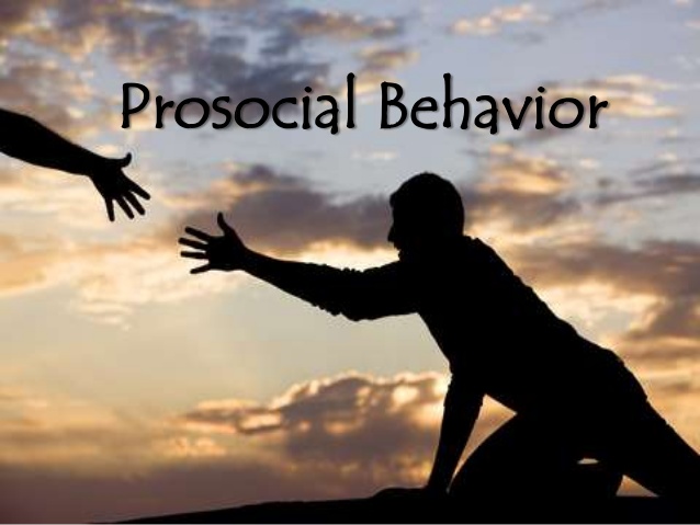
Unfortunately, this view of empathy, as having prosocial implications, is not reflected in the public’s current understanding. An article entitled “Is ‘Empathy’ Really What the Nation Needs?”, from the New York Times, implies that the rise of social networking sites has turned empathy into a “dispassionate approach” that encourages understanding other peoples’ feelings only for self-interest. Various companies advertise through Facebook because they’ve found that emotionally engaging content shares well, and that empathy can prompt real-world action, such as buying their products. Similarly, during the most recent presidential election, the term empathy is said to have been a “buzzword” on Facebook, implying that people should only try to understand others’ motivations well enough to persuade them to vote differently. The public’s current view and use of empathy is quite limited; it has taken the human ability to understand one another and has tried to exploit it. As the attempts to understand others continue to be personal-gain focused, it will be important to research how this egoistic motivation and public’s negative perception of empathy may influence broader social behavior. For example, is it possible that a negative view of empathy could lessen the rewards associated with an empathetic experience, and therefore might decrease the likelihood that an individual would engage in prosocial and empathetic experiences in the future?

References
Hess, A. (2016, November 29). Is ‘Empathy’ Really What the Nation Needs? Retrieved April 23, 2017, from https://www.nytimes.com/2016/11/29/magazine/is-empathy-really-what-the-nation-needs.html
Morelli, S. A., Rameson, L. T., & Lieberman, M. D. (2012). The neural components of empathy: Predicting daily prosocial behavior. Social Cognitive and Affective Neuroscience, 9(1), 39-47. doi:10.1093/scan/nss088
Morelli, S. (2017, March 14). Neural Correlates of Positive Empathy: From Dyads to Social Networks. Lecture presented at the Neuroscience Seminar at Loyola University Chicago, Chicago, IL.

Images

Sunday, April 23, 2017

Prosthetics and the Violin


Dr. Gregory A. Dumanian and colleagues investigated the possibility of reinnervating neural connections for the enhanced use of prosthetic limbs, more specifically, arms. The motivation behind this research resulted from the lack of prosthetic movement due to physical and technological limitations. In one of the studies, a female participant with a left arm amputation went under surgery to isolate and relocate the ulnar, median, distal radial, and musculocutaneous nerves; the same nerves that were severed during the amputation. Before the reinnervation, the nerves were in one large bundle with nothing to pass an action potential to. These free nerve endings may hint to an answer a part of the phantom limb phenomenon. During the surgery, the one bundle was separated into four distinct bundles. The ipsilateral pectoral muscle was separated into four muscles, in order to assign a nerve bundle with a separate pectoral muscle. This process was termed Targeted Muscle Reinnervation (TMR) by Dr. Dumanian. In his presentation, he demonstrated the results of TMR in a video comparing the movement of the same prosthetic before and after the surgery. The difference in fluid motion was astonishing, the results showed a promising future in this field of research. The difference in the two movements was the ability to simultaneously move the arm in multiple directions. For instance, before the surgery, the prosthetic was limited to movement in a vertical, horizontal, or diagonal direction at once. After the surgery, the arm was able to move in circular motions if the patient wished. Obviously, this closely resembles the fluid motion that natural joints allow in upper limbs.
The reason behind this difference was because of TMR. The separation of the one disconnected bundle into four bundles allowed the participant to isolate desired movements, which made them “smarter” signals as Dr. Dumanian described them. One could imagine this like a joystick on a video game controller, one input results in one direction of movement, but a combination of joysticks adds more dimensions into a single movement, i.e. bending at the elbow while rotating the arm. In such a manner, the prosthetic sensors have more input signals from the participant to better interpret the desired combination of motions. Of course, the ability to execute complex motions is the motivating force behind prosthetic research. This is so normal arm function can be restored to the patient. Recently, undergraduate students have created a prosthetic arm for a little girl so she can play violin.

In Fairfax, Virginia, undergraduate students of George Mason University were called upon by an alumnus to work on this project. Isabella Nicola, a fifth-grader at a local elementary school, was born without a left hand, and a disformed forearm. Coincidentally, this group of bioengineer majors were on a hunt for a capstone project to complete for graduation, so this was the perfect opportunity to help both parties. The final prosthetic that the group constructed was made by a 3-D printer, weighing at about 11 ounces. She could play the violin much better thanks to the new arm. They even gave her extra attachments so she can grip a handlebar to ride a bike. The technology used in the prosthetic was not completely described, but it is evident that the complexity of it was enough to allow Isabella to play the violin. With advancements like Dr. Dumanian’s findings, complex prosthetics will be much more common than an undergraduate project taken last second. Hopefully, it will be properly funded and promoted to the point that complexity is not an issue.


References:
http://www.sfgate.com/news/education/article/Prosthetic-arm-designed-by-undergrads-lets-girl-11091145.php 
https://www.youtube.com/embed/TJaGzXle8uA?ecver=2

The Scares of Phantom Limb

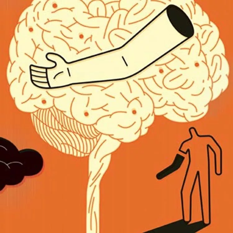
Phantom limb pain is described as a, “pain that feels like it's coming from a body part that's no longer there” (“Phantom pain”). Countless patients have complained about the intense pain experienced when they have had a limb amputated. As of now there has been no cure of treatment for those experiencing phantom limb pain. Many researchers have speculated on what causes this pain in the absence of the limb and some have concluded that the pain is caused by the brain reorganizing neurons and their pathways. This reorganization could be why the brain believes that limb is still attached to the body and why there is so much pain associated with it ("Cause of Phantom Limb Pain in Amputees, and Potential Treatment, Identified”).

Dr. Dumaniam of Northwestern Memorial Hospital has studied how reconnecting key nerves in an amputee’s brain and body can help the patient use their prosthetic to its full extent in his paper, “Targeted reinnervation for enhanced prosthetic arm function in a woman with a proximal amputation: a case study” (Kuiken, et al.). This paper discusses how Dr. Dumaniam reconnected 4 specific nerves in order for the patient to gain full sensory and mobility in their prosthetic so that the patient would be able to live their lives as normally as they did before the amputation. After amputations occur the patient still has a fully functioning nerve, which could be a cause of this painful phantom pain. That is why Dr. Dumaniam has taken the time to figure out which nerves could reconnect and bring back functionality and sensation to the patient. However, the question that does arise is, does the phantom limb pain still occur?
By giving the patient a fully functioning prosthetic does this eliminate the phantom pain because the brain now believes that there is a limb present? Or is it possible that the patient’s brain sees this prosthetic and treats it as a foreign object and reject the entire prosthetic? In a study done in New Zealand, researchers listed some possible reasons why amputees could reject their prosthetic. These reasons include, “development of one handedness which removes the functional need for the prosthesis, lack of sufficient training or skill in using the prosthesis, poor comfort of the prosthesis, the unnatural look or profile of the prosthesis, and the reactions which the wearer gets from other people” (Burrough). These reasons could cause the patient, even more pain alongside the phantom limb pain.

Giving patients fully functioning prosthetics, as Dr. Dumaniam is doing, is an enormous step in the right direction to help amputees live a normal and pain free life. However, phantom limb pain is a serious and intense discomfort to the hundreds and thousands of patients dealing with it everyday. With more research on this pain, hopefully those who have gone through amputations will be able to live their lives as they did before.


Sources
Burrough, Stephen F., and Judith A. Brook. "Patterns of Acceptance and Rejection of Upper Limb Prostheses." Patterns of Acceptance and Rejection of Upper Limb Prostheses | O&P Virtual Library. American Orthotic & Prosthetic Association, n.d. Web. 23 Apr. 2017.
"Cause of Phantom Limb Pain in Amputees, and Potential Treatment, Identified." University of Cambridge. Uni, 27 Oct. 2016. Web. 23 Apr. 2017.
Kuiken, Todd A., Laura A. Miller, Robert D. Lipschutz, Blair A. Lock, Kathy Stubblefield, Paul D. Marasco, Ping Zhou, and Gregory A. Dumanian. "Targeted Reinnervation for Enhanced Prosthetic Arm Function in a Woman with a Proximal Amputation: A Case Study." The Lancet 369.9559 (2007): 371-80. Web.
"Phantom Pain." Mayo Clinic. Mayo Foundation for Medical Education and Research, 03 Dec. 2014. Web. 23 Apr. 2017.
Images
http://neurosciencenews.com/phantom-limb-pain-tms-4830/
http://www.ivketamine.com/phantom-limb/


Wednesday, April 19, 2017

Bionic Prosthetics Using "Smart" Signals


Limb amputations can be extremely debilitating, especially to those who lose an arm above the shoulder. If you think about it, the majority of tasks you complete each and every day revolve around the use of your arm and hands. Without this vital limb, most jobs become overly arduous and some become impossible. This was the case for Les Baugh, who lost both arms at the shoulder due to an electrical accident when he was younger. Despite his devastating injury, Baugh might now able to perform daily life tasks thanks to a new prosthetic arm that is controlled by his thoughts.


In a presentation on Targeted Muscle Reinnervation (TMR), Dr. Gregory Dumanian explained the use of TMR in creating new prosthetics for amputee patients. The big problems of previous models of prosthetics for upper limbs is that those prosthetics can only move one joint at a time and the prosthetic is controlled by the wrong muscle signals.  These prosthetics lack adequate control methods because the proximal muscles used to generate motions of the prosthetic hand and wrist are not normally used to direct those movements.


In order to solve that problem, Dumanian integrated TMR into a new prosthetic limb. Even if an arm is lost, the main nerves in the arm above the injury are still attached to the brain. What he did not yet know was how to utilize those signals to create a “smarter” prosthetic. With knowledge that muscle signals are 100 times stronger than nerve signals, Dumanian was able to create a technique to both utilize the nerves in the arm and the muscle signal generated by the innervation of the arm nerves.


Motor nerves are the nerves that either contract or relax our muscles during coordinated movement controlled by the brain. When a motor nerve is cut, the nerve will search for a receptor or a muscle to innervate. To take advantage of that property, Dumanian took those nerves in the amputated arm and connected them to another muscle that is not normally used, such as the pectoral chest muscle. Connecting these nerves to the new muscle provides a new muscle to be contracted by the signal starting at the brain. From there, he placed electrodes over the newly innervated muscle to detect the “correct” signal. In order to move the hand or wrist, the brain sends the same signals as if the arm were still attached. The signal is instead sent to the newly innervated chest muscle, which contracts. When the muscle contracts, the computerized prosthetic detects the signal pattern and initiates movement of the whole limb, hand, or wrist. These movements are synchronized and executed with as much conscious effort as a person without an amputation. Through TMR and pattern recognition of the signals generated by the chest muscle, this new prosthetic can intuitively “read minds” of those with these inventions. 


With this new procedure and a robotic arm with 26 joints, Mr. Baugh can use his new Modular Prosthetic Limbs (MLPs) to perform daily tasks that do not require proximal muscle use. This new technique and prosthetic now allows for intuitive motion using “smart” signals to generate movement, broadening the potential for dexterity of prosthetic arms and hands. It also points out the greater need for “neural-machine interfaces” to provide sensory feedback to a person with an amputation and allow that person to interact with their environment. However, there is one issue with the MLP: price of the prosthetic. Currently, the Johns Hopkins University Applied Physics Lab is in contact with industry partners to investigate commercial options to make it more affordable for those with upper limb amputations. 


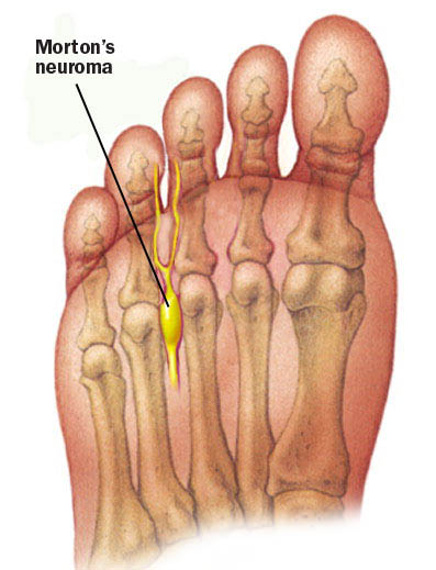
Another interesting feature of Dumanian’s presentation was the discussion about phantom limb pain. When a sensory nerve is cut, neuromas are likely to ensue. A neuroma is the swelling of the nerves or growth of the nerve tissue, which can become very painful. As such, neuromas are very common in patients with amputations. Dumanian found that when TMR and targeted sensory reinnervation (TSR) were implemented to amputee patients, their phantom limb pain prevalence decreased. TSR is similar to TMR because the afferent nerves are connected to a different skin surface. After noticing this correlation, Dumanian began implementing TSR to patients without amputations, but with painful neuromas. He found significant improvements in pain for those patients with Morton’s neuromas and neuromas located in other parts of the body.


Sources
Kuiken, Todd A., Laura A. Miller, Robert D. Lipschutz, Blair A. Lock, Kathy Stubblefield, Paul D. Marasco, Ping Zhou, and Gregory A. Dumanian. "Targeted Reinnervation for Enhanced Prosthetic Arm Function in a Woman with a Proximal Amputation: A Case Study." The Lancet 369.9559 (2007): 371-80. Web