Saturday, April 28, 2018

Two Hit Treatment to Combat Alzheimer's Disease

Two Hit Treatment to Combat Alzheimer's Disease
By Shana Ward 
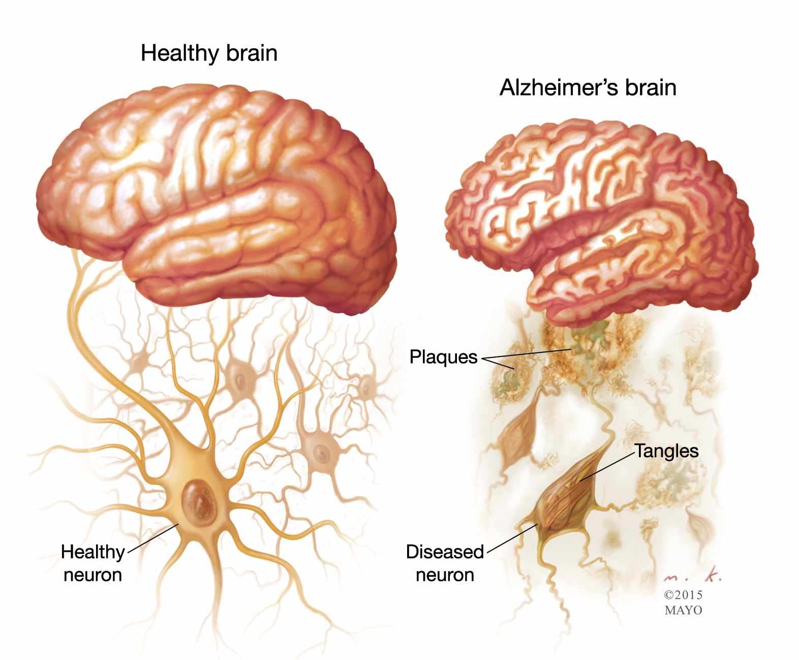


Alzheimer's Disease is one of the top 10 leading causes of death and yet it cannot be prevented, cured, or effectively treated. Dr. Roberto Fernandez gave a talk where he spoke about two major changes that occur in the brain because of the disease, plaques & tangles, which usually result because of protein irregularities. Amyloid plaques are of great interest to many researchers because it is one of the earliest changes that can be detected in the brain. While amyloid is a normal protein found in the brain, pieces of ß-amyloid tend to clump up due to irregular cleavage of the amyloid protein which causes plaque build-ups. 

Recently, there was a study done by Baylor College of Medicine where two approaches to reduce ß-amyloid deposits were at the same time rather than either therapy alone in mice. Jankowsky and colleagues were able to show previously that two treatments that complemented each other worked best to not only reduce the growth of the ß-amyloid plaques but to also clear the plaques that were already present in the brain. With the current study, Jankowsky and colleagues seemed to see real benefits from the dual-treatment through actual improvements in spatial navigation and memory. 

The two treatments included a mouse with genetic modifications that made it able to stop production of the ß-amyloid in the first place and the other treatment included antibodies that bound to ß-amyloid in order to elicit its degradation. Once the mice had undergone both treatments, the researchers were able to see not only reduced plaque levels but also improvements in key cognitive functions like spatial learning and memory very similar to levels that they observed in healthy mice that didn't have the plaques. 

The results of this study could have potentially large implications for people as related to Dr. Fernandez's talk. His talk focused on the visual system since visuospatial symptoms are such common beginning symptoms for AD patients. He talked about areas being affected by Alzheimer's that were similar to areas that are typically activated in a typical visuospatial task. 

Even though the study is implicated in mice, if a similar treatment could be done for humans that improved visual learning, then that would have wide-ranging effects on people with AD who have trouble with visuospatial learning that could impair their driving and other life functions. Hopefully, two hit treatments will be utilized when treating human patients in the future that better the lives of those who have AD. 


https://www.sciencedaily.com/releases/2018/04/180416121539.htm

External Light Effects Circadian Rhythms

It is becoming more evident that humans run on circadian rhythm cycles, which control our sleep-awake routines. One thing that has been shown to effect these defined cycles is the presence or absence of bright light. Researchers have now discovered, using mice as an animal model, that bright lights not only simply affect these rhythms, but can have physical, lasting effects on the body.

Eliane Lucassen and Johanna Meijer of Leiden University Medical Center in the Netherlands have produced pressing evidence that artificial, bright lights can prematurely age mice and cause various health problems, including bone-density loss, skeletal-muscle weakness, and inflammation. While previous studies have been done to hint at bright lights' effects on physical health, direct evidence was not clear.

Lucassen and Meijer implanted electrodes in mice brains to the areas associated with the circadian rhythm cycles and housed these mice in brightly lit cages for 24 weeks. Their discovers showed that neuronal patterns had shifted irregularly, which is a clear association with what happens in aging brains, and led to the mice adopting a roughly 25.5-hour day, rather than a typical 24-hour day. The researchers sought out these experiments to observe the affects humans in busy cities may experience on a daily basis, as roughly 2/3 of the world's population is exposed to light at night. 

These findings coincide with Dr. Cavanaugh's presentation on the circadian rhythm clock that is biologically innate in fruit flies. Dr. Cavanaugh explained the 4 endogenous genes--Clock, Cycle, Period, and Timeless--that are associated with the natural circadian rhythm. The experiments done in Dr. Cavanaugh's lab sought to determine if dimly lit or completely dark environments had an effect on the flies' natural sleeping cycles. He measured the amount of movement the flies had in a tube via infrared beams and found that in constant darkness, the flies did indeed move less, and therefore slept more in this environment, as compared to an environment that stimulated a typical 12-hour light period followed by a 12-hour dark period. 

Together, Meijer's and Dr. Cavanaugh's data conclude that environmental cues of light and darkness can alter the natural circadian rhythms cycles across animal models, and furthermore can have physical health consequences on said models. This could greatly impact the way in which physicians can classify risk factors when considering ailments such as cardiovascular disease, osteoporosis, cancer, and so much more. 


Reference: https://www.scientificamerican.com/article/bright-light-speeds-up-aging-in-mice/
 

Navigating Through Alzheimer's Disease


            Dr. Fernandez explored the navigational impairments of Alzheimer's Disease (AD). Being one of the most prevalent disease for the elderly, it requires attention in order to find ways to cure the problem. Dr. Fernandez looked at the visual processing of optic flow to understand whether there is a higher threshold which leads to navigational impairments. Event related potentials were measured when the participants were subject to driving and navigation. It was clear that patients with early AD has diminishing response amplitudes.
McLachlan and his colleagues looked at AD in terms of memory and a potential threshold effect which may be present. In their experiment, they had three groups: young health individuals, older healthy individuals, and individuals who were diagnosed with AD. Each group was read sentences at one point and then presented more sentences either 1-2 hours and 4-14 days later. They were asked whether the sentences they were being presented were the same as the sentences they had read initially. The results suggested that both the young and older healthy individuals were mainly able to recognize the sentences if they were similar, though the patients with AD were not able to even for short delays. They tested this by looking at the speed of reading the sentence. It has been previously shown how if something is retained, one is able to read it faster.
This experiment was similar to that of Dr. Fernandez’s experiment in that it was measuring the delay patients with AD had when presented a stimulus. To improve the study, McLachlan and his colleagues could have considered looking at ERPs when the participants were given the sentences. This way, they could see what part of the brain was being stimulated initially when the sentences were in working memory and what parts of the brain showed activity when they were actively trying to recall. Perhaps there exist a threshold effect in that the memory of the sentences exists in the AD patients, but the connection is not strong enough for it to be verbalized consciously.
To really get to the cause of the disease, it is important to consider its effects. Patients with AD often have their cortex shrivel, the hippocampus shrinks, and the ventricles become enlarged. Ultimately, the problem boils down to the formation of plaques between the nerve cells. If we can understand the proteins involved in the formation of these plaques, we might be able to use CRISPR to knockout the gene which is resulting in the plaques.
Research in this field is essential as we strive to advance humanity. Because it is a growing problem, it is important to look at the different possible mechanisms of AD in hopes that we find a way to cure it. Currently we cannot cure this neurodegenerative disorder, but we can only down its progression. It is fascinating how one small protein can potentially be the difference between having AD and not having it. Surely, one day we will be able to conquer this disease as well!

Friday, April 27, 2018

The Impact of Caffeine on Hippocampal Long-Term Potentiation


The Impact of Caffeine on Hippocampal Long-Term Potentiation
Daniel Jabr

Dr. Joel Voss of the Feinberg School of Medicine at Northwestern University presented a talk showing how transcranial stimulation to the brain resulted in an enhancement of precision memory recollection.
Specifically, Dr. Voss investigated a particular region of the Hippocampus, the hippocampal posterior-medial (HPM) network, and showed that several days of electromagnetic stimulation facilitated increased recollection of specific, random spatial stimuli, meaning those who received stimulation showed an increased ability to recall the position of random visual stimuli. This finding establishes the HPM to be involved in spatial recollection and recollection of specific visual stimuli.
Dr. Voss further implicates the hippocampus in memory processes and shows that particular regions of the hippocampus can lead to preferential recollection of specific stimuli. The hippocampus has long been known as the major neural substrate for memories. To current knowledge, long-term potentiation (LTP) is the cellular mechanism underlying memory formation. 2 regions in particular of the hippocampus, the dentate gyrus (DG) and the Cornu Ammonis 1 (CA1) regions have been strongly shown to undergo LTP. LTP occurs when neurons of the hippocampus (DG or CA1) experience a tetanus electrical stimulation, that is, a very high frequency stimulation, this causes the synapses between the neurons to strengthen. This strengthening is due to the increased number of glutamate receptors in the post-synaptic cell after tetanus stimulation, meaning the post-synaptic cell will be hypersensitive to the release of neurotransmitter by the presynaptic cell. This strengthening of the synapse by means of increased receptors has been shown to persist (Malenka et al. 2004) with time, as the concentration of the receptors is maintained by the cell due to increased gene expression, yielding increased synthesis of the glutamate receptor which localizes to the cell membrane strengthening the synapse and encoding memories.
Furthermore, the effects of drugs on the nervous system is well documented, with one drug in particular being extremely widespread and is consumed daily around the world. Caffeine is a known stimulant of the central nervous system, increasing alertness and decreasing drowsiness (Mclellan et al. 2016). Does caffeine intake have an effect on LTP?
Blaise et al. (2018) investigated the role of caffeine on hippocampal LTP in rats.10-week-old rats were given access to a reservoir of water for 3 weeks, where the experimental group rats had 1g/L injection of caffeine into their drinking water and the control group had access to only normal tap water, without caffeine. After 3 weeks, the rats in both groups received tetanus stimulation by electrodes projecting to the CA1 region of the hippocampus to induce LTP, while LTP induction in the neurons of the CA1 was measured by recording electrodes. Blaise et al. reported that rats treated with caffeine showed significantly reduced levels of LTP induction with respect to control rats that did not receive caffeine. Hence, those rats that drank caffeine showed less potentiation of the hippocampal neurons closely linked to memory recollection.
These results show that caffeine may negatively impact memory recollection. The researches did not conclude what mechanism may have caused this depression in LTP induction. However, this drop in LTP may be due to caffeine promoting alertness and decreasing sleep in the rats, where the decrease of LTP induction in the neurons is due to the lack of sleep.
Hence, caffeine may result in decreased memory recollection by interfering with LTP in the hippocampus. As caffeine is the most consumed drug in the world, this is a particularly interesting finding, and suggests that extended periods of caffeine consumption may be correlated with decreased LTP in hippocampal neurons. As such, drinking caffeine might result in poorer performance on cognitive tasks and memory retrieval, not something that society today would be excited to here as caffeine is the most consumed drug that prepares people, from students to construction workers, for the day. Furthermore, caffeine interferes with the normal sleep-wake cycle and inhibits sleep to some extent. Thus, caffeine might not be the wonder-drug that it seems, and may lead to decreased memory recollection.

Link to journal article: https://www.ncbi.nlm.nih.gov/pmc/articles/PMC5840440/

References:
Malenka, RC, Bear, MF. 2004. LTP and LTD: An Embarrassment of Riches. Neuron 44:5-21
McLellan, TM, Caldwell, JA, Lieberman HR. 2016. A review of caffeine's effects on cognitive, physical and occupational performance. Neurosci. Biobehav. Rev. 71:294–312. 

Blaise, JH, Park, JE, Bellas, NJ, Gitchell, TM, Phan, V. 2018. Caffeine consumption disrupts hippocampal long-term potentiation in freely behaving rats. Physiological Reports 6(5): e13632