
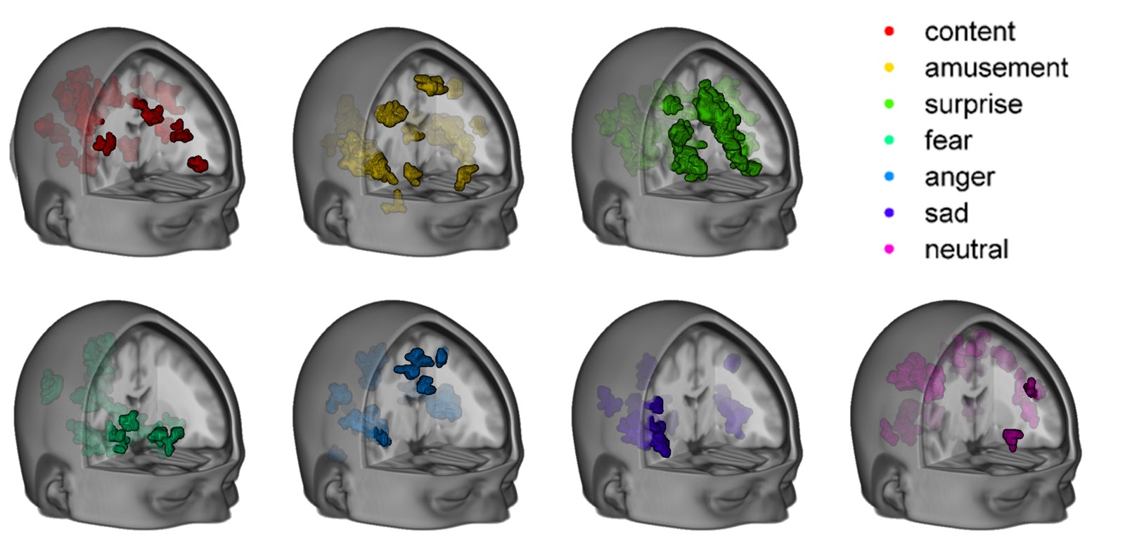
Veronique Greenwood of the Scientific American proposes a question about the cognitive nature of emotions, and whether brain region activation varies among different individuals who have their own subjective experiences of emotions. The author then explores this subject by mentioning recent studies that have employed Functional Magnetic Resonance Imaging (FMRI) scans to pinpoint the specific locations of the brain that are activated when we experience certain emotions such as fear, happiness, sadness, or anger.
Greenwood mentions a study published by PLOS Biology in September 2016 in which researchers from Duke University averaged FMRI results of multiple subjects who had underwent specific emotional provocations (achieved by having participants watch dramatic movie clips or listen to key-words) and retroactively predicted a certain emotion by comparing an activated brain scan to an neutral brain scan. The researchers found that they were successful in guessing the emotion about 75% of the time. However, Greenwood points out that the studies she has mentioned may not be reliable across different populations of individuals because, as professor Lisa Feldman Barrett of Northeastern University observes, the patterns taken from these studies have not been successfully replicated in different groups of individuals, as the results of these studies are based on statistical summaries and not the unique localizations of brain activity that occur when someone has their own subjective emotional experience.
MRI relies on the magnetic properties of organic tissue to map out structures of varying densities, producing detailed and clear images of our bodies through the mapping of proton movements in response to the pull of the strong magnetic field of an MRI machine. Functional MRI employs the technology of MRI to measure time-dependent fluctuations in the brain’s oxygenation levels in response to stimuli. The studies mentioned in Greenwood’s article employed this technique to measure individuals’ Blood-Oxygenation Level Dependent (BOLD) increases in specific regions in response to emotion-evoking stimuli such as dramatic movie clips or certain key-words. However, as the author of the article points out, different brain region groups may be activated in different people who are experiencing the same emotion, as the evocation of emotion is not always dependent on a concrete stimuli, as it also results from the wandering of our thoughts and memories.
A study conducted by Sun et. al. in 2004 (taken from our Cognitive Neuroscience textbook) studies the functional connectivity, (the idea that activation in one brain area is related to activation changes in another area). This idea is exemplified in the dispersed BOLD signals of multiple brain regions when differing emotions are elicited in the studies mentioned by Greenwood. Additionally, in a 1999 study conducted by Deilbert et. al., tactile object recognition led to the BOLD activation of the visual cortex, showing that touch is associated with mental visualization of an object even when a subject’s eyes are closed. However the difference between emotion-dependent brain region activation and action- or touch-dependent activation is that emotion is the result of more complex and subjective neural networks that are constructed based on individual memories retrieved and activated by the hippocampus. Though certain brain regions are synonymous with certain emotions (i.e. the amygdala is often known as our fear center, though it also controls the expression of anger and sadness), the neural networks between different brain regions are still likely based on each person’s specific neural pathways between brain regions that are activated when they experience a specific emotion.
article: https://www.scientificamerican.com/article/can-you-tell-someones-emotional-state-from-an-mri/
No comments:
Post a Comment